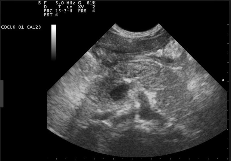
Ultrasound Artifacts 5: Image Optimization

Ultrasound artifacts, which are becoming increasingly rare as technology improves, can still lead to critical errors in interpretation if they are not recognized and dealt with appropriately. A proper understanding of how they are created allows the echocardiographer to recognize, and work around these potentially hazardous pitfalls. This course covers recommendations for image optimization, including use of harmonic imaging and system gain, dynamic range, and variance mapping, among others.
(NOTE: This material is best understood after completing The Physics of Ultrasound series first.)
In this course, you will learn:
Recommendations for image optimization, including:
- gastric decompression
- use of harmonic imaging and system gain
- finding the optimum depth, width, zoom, and focus
- dynamic range
- color gain
- variance mapping
Method and medium:
Learners participate in the interactive learning modules by correctly answering multiple choice questions dispersed throughout. Learners will be prompted to try again if a question is answered incorrectly.
The course will open in a new tab – to exit the course, simply close that tab.
Estimated time to Complete: 30 minutes
Credit/contact hours: .5
Expiration date: March 7, 2021
Course published March 8, 2018
