Aortography And Ventriculography
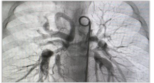
Aortography and ventriculography are more specialized imaging techniques used in the cardiac catheterization laboratory for specific diagnoses and for specific indications. Ventriculography is an important diagnostic imaging modality to assess left ventricular function, regional wall motion abnormalities, severity of mitral regurgitation, certain congenital and shunt conditions such as ventricular septal defect.
During acute MI, ventriculography can be useful to determine an infarct zone or infarct artery. In some cases, ventriculography may make less common diagnoses such as ventricular aneurysms, Takotsubo cardiomyopathy, or ventricular diverticulae. Aortography is commonly used to make diagnoses in aortic valve disease as well as to define aortic pathology such as aneurysms. Occasionally, the location of venous bypass grafts are best determined by aortography. Proper technique and interpretation are always required.
In this course, you will learn:
- To define the diagnoses that are made by ascending aortic aortography.
- To correlate the optimal imaging views used for aortography and ventriculography to specific disease states.
- To characterize the severity of aortic regurgitation based on angiographic and hemodynamic information.
- To identify and characterize ejection fraction, regional wall motion abnormalities, aneurysms, and less common conditions such as Takotsubo cardiomyopathy or ventricular septal defect by left ventriculography.
Method and medium:
Learners participate in the interactive learning modules by correctly answering multiple choice questions dispersed throughout. Learners will be prompted to try again if a question is answered incorrectly.
The course will open in a new tab – to exit the course, simply close that tab.
Estimated time to Complete: 40 minutes
Credit/contact hours: .75
Expiration date: March 7, 2021
Course published March 8, 2018